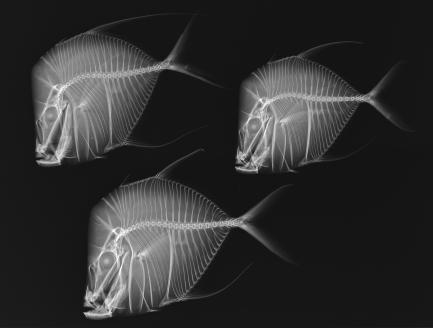
Lookdown fish (Selene vomer) specimens in an x-ray exposurefrom “X-ray Vision: Fish Inside Out,” an exhibition from the Smithsonian’s National Museum of Natural History and the Smithsonian Institution Traveling Exhibition Service (SITES).
Fish are vertebrates—animals with backbones—and have bodies supported by a bony skeleton. Variations in the skeleton, such as the number of vertebrae or the position of fins, are documented with X-rays. The Smithsonian’s national collection of these fish X-rays represents more than 70 percent of the world’s fish specimens and is the largest and most diverse collection of its kind in the world. Although the X-rays featured in the national collection were made for research purposes, the strikingly elegant images demonstrate the natural union of science and art and are a visual retelling of the evolution of fish. “X-ray Vision: Fish Inside Out,” an exhibition from the Smithsonian’s National Museum of Natural History and the Smithsonian Institution Traveling Exhibition Service (SITES), will showcase these dramatic prints exposing the inner workings of the fish.
“X-ray Vision” will premiere at the Yale Peabody Museum of Natural History in New Haven, Conn., July 2 and will be on view until Jan. 8, 2012. The exhibition will then travel around the country on a 10-city national tour through 2015.
The exhibition features 40 black-and-white digital prints of several different specimens of fish. Arranged in evolutionary sequence, these X-rays give a tour through the long stream of fish evolution. The X-rays have allowed Smithsonian and other scientists to study the skeleton of a fish without altering the sampling, making it easier for scientists to build a comprehensive picture of fish diversity.
Curators of the exhibition, Lynne Parenti and Sandra Raredon, have worked in the Division of Fishes at the National Museum of Natural History collecting thousands of X-rays of fish specimens to help ichthyologists understand and document the diversity of fishes. Rare or unique specimens make particularly interesting and informative images. X-rays may also reveal other details of natural history: undigested food or prey in the gut might reveal to an ichthyologist what a fish had for its last meal. To make comparisons easier, radiographers X-ray one fish per frame—with each one facing left—but they will prepare shots of several fish if a scientist wants to compare a group.
“X-ray Vision: Fish Inside Out” is inspired by the book Icthyo: The Architecture of Fish: X-Rays from the Smithsonian Institution (Chronicle Books, 2008) by Daniel Pauly, Lynne Parenti and Jean-Michel Cousteau.
The Smithsonian’s National Museum of Natural History, located at 10th Street and Constitution Avenue N.W. in Washington, D.C., welcomes more than 6 million visitors annually. The museum is open daily from 10 a.m. to 7:30 p.m. every day during the summer from March 25 through Sept. 4. Visit www.mnh.si.edu for early closure on select days throughout the summer. Admission is free. For further information, call (202) 633-1000, TTY (202) 633-5285 or visit the museum’s website at www.mnh.si.edu.
SITES has been sharing the wealth of Smithsonian collections and research programs with millions of people outside Washington, D.C., for almost 60 years. SITES connects Americans to their shared cultural heritage through a wide range of exhibitions about art, science and history, which are shown wherever people live, work and play. Exhibition descriptions and tour schedules are available at www.sites.si.edu.
# # #
SI-152-2011